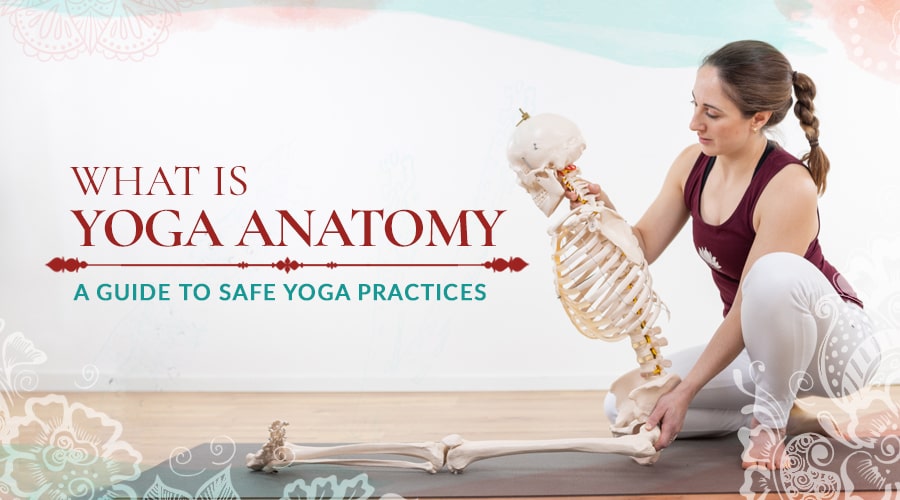
Anatomy knowledge will help you practice safe yoga. If you’re a yoga teacher, knowing yoga anatomy will help you teach safe, effective and more enjoyable yoga to your students. Anatomy helps explain the individual variations in progress and the appearance of yoga poses.
What is Yoga Anatomy
Anatomy is a field of biological science studying the structure of the body. Anatomical science typically views the body as a composite of many separate parts and searches out the details of each part. Although this might not be a holistic way to view the body, the information can be very useful when applied within a more holistic approach. Yoga anatomy encompasses the science of anatomy as it applies to the practice of yoga, and particularly the practice of yoga asana.
Anatomy or physiology?
Anatomy refers to the structure of the body and the physical relationship between body parts. Physiology refers to the function of body parts and the functional relationships between them. Consider the nervous system: how and why it transmits messages belongs to the science of physiology. Where the nerves are located in their paths around joints or through bones is the science of anatomy.
In practice, anatomy is often about bones and muscles, while physiology is often about organs.
What should yoga teachers know about yoga anatomy?
Learning about anatomy is a process. If you try to pack all the information into your brain too quickly, it will seem ridiculously complicated and frustrating. A better strategy is to read and absorb the information that makes sense to you… and then come back later to read the information again. You will find your understanding growing over time.
Anatomy is a huge scientific field. You could spend years studying it, earn a master’s degree and a handful of scientific awards, and still be quite certain there’s more to learn about the structures of the body.
Fortunately, you don’t need to be an expert or a scientist to have a useful working knowledge of anatomy for yoga. You just need to be curious, interested, and willing to learn.

Teaching Yoga Anatomy
The language of anatomy
In yoga, the traditional language is Sanskrit. Some people learn the Sanskrit names for poses and other elements of yoga; other people learn versions of the words and names in their own language. Something similar is true for anatomy.
The traditional language of science is Latin, with a little ancient Greek mixed in. Having a common language allowed scientists from different countries to communicate and share their knowledge.
You can develop a good understanding of anatomy for yoga without learning all the formal terms, just like you can practice a Seated Forward Bend without remembering it’s also called Paschimottanasana.
The Most Fundamental Principle of Yoga Anatomy: Everybody’s Anatomy is Different
This is an important and often-forgotten element of anatomy. It’s extremely important information for practitioners and teachers of yoga.
We know people can be taller or shorter, and some have commonly recognized differences like broader shoulders or larger feet. However, normal anatomical differences can also change the proportions of the body, and this has a direct effect on practicing yoga asana.
Two people might be the same height, but one might have a short torso and longer legs. The other might have a longer torso and shorter legs. When the first person practices Child’s Pose, their knees will be near their shoulders. Their chin tucks towards their neck and their forehead rests on the floor. When the second person practices Child’s Pose, their knees will be near their lower ribs. Their shoulders rest closer to the floor. This places their face flatter to the floor, prevents the chin tucking in, and results in a position nose-first into the floor instead of more comfortably resting on their forehead.

One expression of Child’s Pose
Differences like this apply to a great number of poses. Anatomical differences can include leg or arm length, torso length, and differences in muscle bulk that can alter the movement available. A person with muscular thighs will have more difficulty twining their legs together in Eagle Pose (Garudasana), or getting their knees stacked in Shoelace Pose (Gomukhasana) - even more so if they are also short. A person with longer and more slender legs will find those poses easier to access, even if their joints are no more flexible than the muscular person’s.
Another very common anatomical difference can affect the amount of movement available at a joint. The joint with the most obvious variability is the hip, a ball-and-socket shaped joint connecting the thigh to the pelvis. The bony shape of the hip socket in the pelvis, and the shape and orientation of the “ball” at the top of the leg can vary greatly - while still being normal and healthy. This means some people will have greater or lesser movement in one or more directions at the hip. Practice and flexibility training can increase the movement allowed by muscles and ligaments, but they will not change the bones of a joint. Trying to force movement onto a joint limited by bone is likely to cause injury, possibly even fractures.
All You Need to Know About Yoga Anatomy
The Skeleton

The human skeleton
The human body is built from approximately 206 bones (approximate because that too can vary between people). About a quarter of these bones are in the hands and feet, which typically contain 53 bones altogether.
One of the main reasons for becoming familiar with the bones of the body is their role in supporting our muscles and movements. Knowing the names of the major bones makes discussing or studying movements much easier.
Places where one bone meets with another bone are called joints, and these are even more important when learning about movement. Each unique human has around 300 joints.
The skull, spine and ribcage together are called the axial skeleton. They form a base for our more mobile limbs. Attaching to this base, the pelvis, shoulder girdle and limbs are collectively called the appendicular skeleton.
In yoga, movement in the axial skeleton occurs in the spine and ribcage. The spine has three sections which differ in structure and movement. They are the cervical spine (neck), thoracic spine (upper back) and lumbar spine (lower back). During yoga asana, the three regions of the spine work together to produce the required range of motion.
The anatomy of the spine
The neck or cervical spine
There are seven vertebrae in the neck or cervical spine. Most of the flexion and extension (forward and backward bend) in the cervical spine comes from the top joint where the skull meets the first vertebra. Most of the neck’s rotation comes from the joint between the first vertebra and the second vertebra. The remaining joints in the cervical spine allow movement in multiple directions, adding up to greater flexion forward, backward and sideways than any other area of the spine.
In joints, greater flexibility means greater vulnerability. Movement at the neck should never be forced.
While most of the spine has a shock-absorbing pad called an inter-vertebral disc in between each vertebra, the discs of the cervical spine are the smallest and thinnest. There are no discs between the skull, the first vertebra, and the second vertebra.
Weightbearing through the neck (eg. headstands and shoulderstands) should be approached carefully with special attention to bodily feedback and appropriately gradual progression of practices. The spine is designed to bear the body’s weight through the large strong vertebrae and thick resilient discs of the lower back, but with care and common sense it’s possible to safely practice inversions that put the body’s weight through the small vertebra of the neck.
(For more tips on safe headstands, see How to do Headstand)
The ribcage and thoracic spine (upper back)
The ribcage consists of 12 (rarely 13) pairs of ribs that connect to the spine at the back. At the front of the ribcage, the upper ribs connect to each other and to the breastbone or sternum. The lowest two ribs are much shorter and don’t connect at the front. The ribcage has two primary roles:
- protecting the thoracic organs
- Aiding respiration - the lungs don’t move by themselves, they are expanded and contracted when our muscles move our ribs and diaphragm.
The spinal bones of the upper back are called thoracic vertebrae. There are twelve, each with a bony outcrop to each side for the ribs to attach to. Movement in the thoracic spine is limited by the joints between the vertebrae, and the bony shape required for the rib attachments. The thoracic spine has less flexion and extension (bend forwards and backwards) than the neck or lower back. On the other hand, it has more rotation (twist) than the rest of the spine.
Asanas that encourage mobility in the thoracic spine and ribcage can improve breathing and reduce back pain, arm and shoulder pain and headaches.
The lower back or lumbar spine
There are five vertebrae in the lower part of the spine. They are the largest vertebrae, separated by thick inter-vertebral discs that support the upper parts of the spine and the weight of the upper body.
The lumbar spine has its greatest range of movement in backward bending (extension), and also allows forwards and side bending and a small degree of rotation.
Although the lumbar spine is strong and resilient, it should not be forced beyond its natural range. Forward bends are common and valuable asanas in yoga practice, so students and teachers should learn how to practice forward bends safely.

Spine and pelvis
Spine safety tips for yoga
Spinal cord: the spine forms a bony protective tunnel for the spinal cord. At each joint between vertebra, there is a channel for a bundle of nerves to connect the spinal cord to the rest of the body. Injuries to the spinal bones or joints can directly injure these nerves, or cause swelling which in turn puts pressure on the nerves.
The spine’s direct relationship to the nerves gives very good reason to treat the spine with respect and listen to feedback from the body. Forcing too much movement through the spinal joints can cause nerve symptoms like pain, tingling or numbness in areas the nerves travel to. Sensation in the hand might come from pressure in the upper back; sensation in the leg or foot might come from pressure in the lower back. Causing these symptoms is damaging to the nerve and counterproductive to healthy yoga practice. It’s important to allow flexibility to develop gradually in each individual, and respect their anatomical limits.
Spinal curves: Each section of the spine is naturally curved. The curve rolls in at the neck, out over the upper back, and in again at the lower back. This rhythm even continues into the pelvis, which forms another outward curve below the lower spine. These curves are important for balance, weight distribution and upright posture. They are natural and normal, vary in degree between individuals, and there is no need to try to ‘correct’ them. If you or your student appears to have abnormal curvature in the spine, there are many possible explanations. A medical professional should be consulted for guidance and safety.
Spinal mobility: Keep in mind that most rotation should come from the upper back. The neck is highly mobile but should not be pushed into vulnerable extreme ranges of movement. If an asana is difficult to achieve, take note of where movement seems to be blocked. It is best to work on increasing range in the restricted area, rather than allowing other areas of the spine to compensate for one inflexible area (with respect for existing injuries/conditions).
Experience authentic Hatha Yoga
Get free access to exclusive guided lessons with master teacher Kalyani Hauswirth-Jain
The pelvis
The pelvis is a bony circle that supports the spine and torso. The large triangular bone under the lumbar spine is the sacrum, and below it sits the small coccyx or tailbone. On each side of the sacrum, the pelvic bones circle around to join at the centre front. The joint at the centre front of the pelvis contributes to stability and should have very little movement. The sacro-iliac joints - where the pelvis joins the sacrum at the back - have a very small degree of movement. It can be helpful to gently encourage movement at these joints. Supta Virasana (Reclining Hero Pose) is intended to mobilise the sacro-iliac joint.
Yoga safety tip - pelvic stability
Care should be taken with pelvic joint mobility because encouraging mobility can easily create instability, which can cause considerable pain either during the practice or in daily life. Pregnant and post-natal women, and hypermobile (overly flexible) people are particularly vulnerable to issues of pelvic instability.
Some poses can also stress the sacro-iliac joints unevenly, which may be unsafe or uncomfortable for people with reduced stability. Weightbearing poses with asymetrical leg positions cause uneven weightbearing through the pelvic joints, particularly if the legs are wide apart.
Joints of the skeleton
Joints come in many shapes to allow a variety of movements. The major movements of the body occur at synovial joints. These joints are characterised by a lubricating fluid called synovial fluid, which coats the cartilage on the end of each bone and facilitates smooth movement of the bones against each other. The joints are held together by a capsule of connective tissue and an assortment of ligaments.
These joints are sorted into types by their shape, which dictates what movement they can allow:
- Hinge joints bend in one direction only (eg. elbow)
- Pivot joints only allow rotation (eg. joint between top two vertebra of the neck)
- Saddle joints allow bending forwards, backwards and sideways (eg. base of thumb)
- Condyloid joints bend in the same directions as saddle joints, with less range (eg. between radius and the carpal bones)
- Plane joints allow a sliding or shearing motion (eg. the sacroiliac joint - back of pelvis)
- Ball and socket joints allow movement in all directions (eg. shoulder and hip)

Knee joint
There are two other types of joints that may be of interest to yoga practitioners.
Fibrous joints don’t look like what most people think of as a joint and include the suture lines of the skull. There are two fibrous joints more relevant to you. In the forearm and lower leg, the bone pairs are bound together by strong connective tissues and fascia, but they allow some movement in cooperation with movement at the wrist and ankle. It is possible to damage these tissues if wrist or ankle movement is forced beyond the person’s safe range of flexibility.
Cartilaginous joints have joint cartilage but no synovial fluid. They may also have a fibrous disc acting as a shock absorber and creating space for movement - these include the intervertebral joints of the spine, as discussed above.
Joints in yoga
- Joints should move only in the directions they were designed to move. Trying to force other movements can cause injuries. It’s important to take care with asana progressions that press joints in multiple directions. Some variations of Pidgeon Pose, for example, require knee rotation which is a very limited movement.
- The same joints are slightly different shapes in different people. This means one person can have a greater range of movement than another person because of the bone shapes they were born with.
- Stretching a joint beyond its normal limits can stretch and weaken the ligaments and connective tissue that are supposed to support and protect the joint.
- Popping and clicking during normal movement does not necessarily indicate injury. It often suggests the joint is a little unstable and might benefit from muscle strengthening around the joint.
- Intentionally ‘cracking’ joints on a frequent basis is not recommended as it can cause wear on the joint surfaces and weaken the supportive tissues around the joint.
- Strong coordinated muscles are a safety net for our joints. Increasing flexibility in a joint should always be teamed with increasing muscle strength and control of the new range of movement, to keep the joint safe.
- Hyperflexion and hyperextension are movements that go beyond the normal limit permitted by a joint. This can result in ligament tear, damage, or dislocations.
- Some people have naturally occurring hypermobility (extra flexibility), but this does not mean their joint is safe in a hyperflexed or hyperextended position. They are likely to be at greater risk of injury because their joints are not protected by connective tissue controlling movement beyond the normal limits.
Yoga anatomy - muscles
There are over 600 muscles in the human body. We do not need to learn all of them to practice or teach yoga.
Some muscles, like the heart and the muscles that propel food along the digestive tract, are not under our conscious control and are called involuntary muscles. The muscles we use intentionally to move about are called voluntary muscles. Some, like the muscles used to breath, have both voluntary and involuntary control.
Each muscle used to move our skeleton has an origin and an insertion: the points each end of the muscle attach to. Each muscle also has an action, which is the body movement that occurs when the muscle contracts. If a muscle crosses a joint, it’s action will be to cause movement in that joint. This simple concept is complicated by several factors:
- Some muscles cross more than one joint.
- Some muscles can cause movement in more than one direction eg bending and rotating the joint.
- Multiple muscles can work in combination to produce a desired movement.
- Muscles contract (shorten) to cause movement, but they can also work while lengthening to control the return path of the movement.
- Often in one yoga pose, multiple movements are taking place at the same joint at the same time.
Yoga anatomy – fascia
Fascia is an extremely important element of human anatomy, and anatomy in yoga. Made up of connective tissues, if forms a web that supports and connects everything in our body. There is fascia under our skin, fascia wrapping our organs and muscles, and fascia forming a fine web around bundles of fibres inside each muscle. Fascial tissue ranges from dense and strong to exquisitely fine.
When the body moves, fascia moves. If fascia is not moved regularly, it starts to bind together, creating stiffness in the body. Fascia also relies on movement to deliver hydration, since it doesn’t have a blood supply. Dehydrated fascia also reduces flexibility and is thought to be the source of many symptoms people think of as old age reducing their mobility.
Fascia is best kept healthy by regular practice of a wide variety of movements and positions – a perfect description of yoga practice. Just like a car seatbelt, fascia will extend during slow movement but slam on the brakes if you push quickly into a stretch. For this reason, slow flowing practices and practices with held poses are good for improving flexibility via the fascia.
Injury Prevention in Yoga
- Take note of the spinal safety and pelvic stability tips above.
- Always warm up at the beginning of a physical yoga practice. Warming up increases the coordination and reaction time of muscles, greatly improving their ability to protect themselves and your joints. It also increases blood flow and prepares muscles for a safe and efficient increase in activity and mobility. Gentle Sun Salutations are an effective warmup included in many yoga practices.
- Remember that each person’s natural alignment in an asana is likely to be different due to variations in bones, joints, strength, fitness, flexibility and other factors. Each person should practice appropriately for their own progress, not a fictional ideal image of a pose. This also applies to the duration a pose is held.
- Progress is unique to individuals. With practice, a person may improve in capacity in many areas including flexibility, cardiovascular fitness and strength of muscles and bones. Progressions should be made carefully and slowly to avoid injury - overuse injuries often appear after weeks of excessive overload rather than during the first overloaded activities. Progressions might be in the length of time a pose is held for, the range of movement used in a pose, or the practice of more complex poses.
- A person who has had a break from physical practice will need to start slowly and build their capacity back up to their previous level. The body does not retain strength, endurance or other capacities if they are not being used regularly.
- Rest is vital for both injury prevention and physical progress. Body tissues are challenged during practice, but it’s during rest that they build and repair to make themselves more able to meet the challenge. Progress is a result of combining challenge and rest. Just like challenge and capacity, rest requirements vary between people. The need for rest is reflected in duration of a practice, time between practices, and number of rest days built into a weekly schedule. It’s also important to get good sleep!
Conclusion
Anatomy gives us a foundation for how each yoga pose affects the body. Based on the physical framework of bones, joints, muscles and fascia, we come to understand that each human body has these things in common, but at the same time each body is unique. Each person’s practice will look different in order to challenge, strengthen and nurture their unique anatomy.
In practicing yoga, which seeks unity, we must recognise these differences. Every person can unite in yoga practice, and be accepted in their unique expression of each asana.
Sources
Hatha Yoga for Teachers & Practitioners - https://www.amazon.co.uk/dp/9082705613
Experience authentic Hatha Yoga
Get free access to exclusive guided lessons with master teacher Kalyani Hauswirth-Jain